![]()
"The study of dental morphology is essential in terms of phylogeny.
Advances in three-dimensional (3D) measurement devices have enabled us
to make 3D images of teeth without destruction of samples. However, raw
fundamental data on tooth shape requires complex equipment and techniques.
An online database of 3D teeth models is therefore indispensable. "
(A. Kato and N. Ohno. (2009) Construction of three-dimensional tooth model
by micro-computed tomography and application for data sharing. Clinical
Oral Investigations 13, 43-46.)
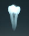
| Tooth | image photo | X-ray photo | OBJ data | STL data | DXF data | QTVR |
| Upper left central incisor (FDI: 21) | 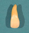 |
 |
ULI1_OBJ.lzh | ULI1_STL.lzh | N/A | |
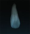
| Upper left lateral incisor (FDI: 22) | 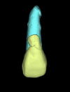 |
(left: rendered 3D model) |
22ULI2_OBJ.lzh | 22ULI2_STL.lzh | N/A | N/A |
| Lower right first premolar (FDI: 44) | 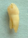 |
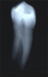 |
44LRP1_OBJ.lzh | 44LRP1_STL.lzh | N/A | N/A |
| Lower left second premolar (FDI: 35) | 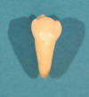 |
 |
N/A | N/A | 35LLP2.lzh | |
| Upper right first molar (FDI: 16) | 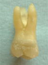 |
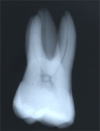 |
16RUM1_OBJ.lzh | 16RUM1_STL.lzh | N/A | N/A |
| Lower right first molar (FDI: 46) | 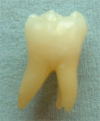 |
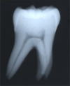 |
46RLM1_OBJ.lzh | 46RLM1_STL.lzh | N/A | N/A |
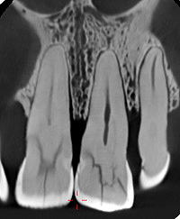
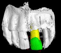
Teeth surrounded Maxillary & Mandibular bone
| CT image | Rendered image | Segmentation data | STL data | |
| Upper left central Incisor (LUI1) & bone |
 |
 |
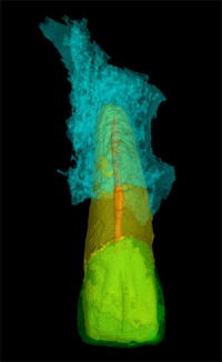 |
Enamel, Dentine, Pulp cavity, Bone On request |